Pain and Temperature Pathway
Sensory neurons in the skin > Dorsal Root Ganglion of the Spinal Cord > Dorsal Horn > Lateral Spinothalamic Tract > Ventral Posteriolateral nucleus of Thalamus > Somatic Sensory area of the Cortex in Postcentral Gyrus (areas 1,2,3). Lesions produce neuralgia and phantom limb pain.
Pressure and Crude Touch Pathway
Sensory neurons in the skin > Dorsal Root Ganglion of the Spinal Cord > Dorsal Horn (one branch ascends ipsilaterally)> Ventral Spinothalamic Tract (most meurons decussate here) > Ventral Posteriolateral nucleus of Thalamus > Somatic Sensory area of the Cortex in Postcentral Gyrus (areas 1,2,3). Partial lesions in the cord produce partial loss of sensation because of dual pathway, higher lesions produce loss of sensation.
Proprioception, Fine Touch, and Vibratory Sense Pathway
Sensory neurons in the skin > Dorsal Root Ganglion of the Spinal Cord > Fasciculus Cuneatus (thoracic and cervical levels) and Fasciculus Gracilis (sacral and lumbar levels) > Nucleus Cuneatus and Nucleus Gracilis of Medulla > Decussation in Medulla > Medial Lemniscus > Midbrain > Ventral Posterolateral Nucleus of Thalamus > Somatic Sensory area of the Cortex in Postcentral Gyrus (areas 1,2,3). Lesions produce astereognosis (loss of ability to recognize objects by touch – key, coin, paperclip test), loss of vibratory sense, loss of two-point tactile discrimination (compass test), loss of proprioception (touch your nose with your eyes closed, Romberg test – swaying while standing erect with both feet together and eyes closed)
Voluntary Muscle Activity Pathway
Motor Cortex in Precentral Gyrus (areas 4,6).> Internal Capsule > Basis Pedunculi Cerebri in Midbrain > Pyramidal Tract > Decussates in Medulla (Ventral Corticospinal Tract contains 10-20% of axons that don’t decussate) > Lateral Corticospinal Tract > Ventral Horn > Ventral Root > Nerve endings in voluntary muscles. See CNS anatomy notes for UMN and LMN lesions.
Vestibular System
Sensory neurons in the inner ear > Vestibular Ganglion > Vestibular Nuclei (Area Acoustica of the floor of the Fourth Ventricle) > Inferior Cerebellar Peduncle > Cerebellum (there’s feedback loop from Cerebellum to Vestibular Nuclei and a pathway to motor neurons via Medial Vestibulospinal Tract, there are also connections to cranial nerves III, IV, VI). Lesions produce disturbances in equilibrium, walking, dizziness, nystagmus (diff. Meniere’s disease).
Cerebellar Pathways
In addition to the Vestibulocerebellar pathway, Cerebellum receives information from muscle, joint and tendon receptors via Ventral Spinocerebellar Tracts and Dorsal Spinocerebellar Tract and from the Cortex (secondary motor areas) via Corticocerebellar fibers connecting at the Pontine Nuclei and decussating in Middle Cerebellar Peduncles; there are feedback loops to the cortex (Primary motor) via Dentorubrothalamic Tract, connecting in the Red Nucleus and Thalamus and to the muscles via Red Nuclei and Rubrospinal Tract. Cerebellar lesions produce Asynergia (loss of coordination and smoothness in motor actions), Dysmetria (inability to judge distances), Adiadochokinesia (inability to perform rapid alternating movements), Intention tremor, Ataxia (abnormal gait, compensatory wide gait), Falling (towards the lesion), Hypotonia or hypertonia of the muscles, Dysphonia (slurred, explosive speech), Nystagmus.
Autonomic Nervous System
Autonomic Nervous System consists of two antagonistic systems supplying the same organs (cardiac muscle, most glands and all smooth muscle): Sympathetic System (fight or flight – increased breathing rate, pulse, blood pressure, blood flow to heart and voluntary muscles, decrease blood flow to skin, decreased peristalsis, dilated pupils, increased sweating, hair “standing”, increased glycogen secretion) and Parasympathetic (relaxation – opposite effect). Activation of one system suppresses the other one. Connect to the Cortex via Hypothalamus. Adrenergic drugs (adrenaline, noradrenaline, and dopamine) mimic sympathetic activity and cholinergic drugs mimic parasympathetic activity, blockers have opposite effect. Lesions in the sympathetic system may produce Horner’s syndrome: Anhydrosis (no sweating), Miosis (fixed small pupil), Ptosis (eyelid drooping), and flushing of one side of the face.
Auditory Pathway
Hair cells in Cochlea > Spiral Ganglion > Auditory part of Cranial Nerve VIII > Ventral and Dorsal Cochlear Nuclei in Pons > part of the fibers decussate at the Trapezoid Body (both ears are represented in both hemispheres) > Superior Olivary Nucleus > Lateral Lemniscus > Nucleus of Inferior Colliculus in Midbrain > Medial Geniculate Body > Auditory Radiations > Auditory center of the cortex (areas 41 and 42).
Olfactory System
Receptor cells in the nasal cavity > Olfactory Bulb > Olfactory Tract > bifurcates into Lateral and Medial Olfactory Tracts > Paraolfactory and Anterior Perforated Areas (some fibers decussate here, both sides are represented in both hemispheres) > Amygdaloid Nucleus > Uncus and some fibers go via Stria Terminalis to Hypothalamus, then Amygdala, Hyppocampus, and Fornix back to Hypothalamus. There are asociated reflex pathways to motor nuclei and thalamus (Mamillotegmental and Dorsal Longitudinal Fasciculi and Mamillothalamic Tract to Thalamus and then Cingulate Gyrus, respectively). Periferal lesions produce Anosmia (loss of smell), amygdala and uncus lesions produce olfactory hallucinations and sometimes epileptic seizures with olfactory aura (Uncinate fits).
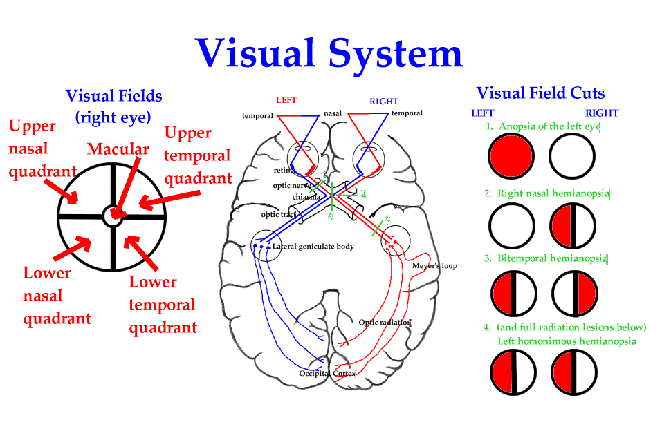
Visual Pathway
Receptor cells in the retina > Optic Nerve > Chiasma (fibers from the opposite visual field decussate here so that opposite visual fields from both eyes are represented in each hemisphere) > Optic Tract > Lateral Geniculate Body > Optic Radiation > Occipital Cortex (information from upper visual field goes to Lingual Gyrus below Calcarine fissure and from lower field to Cuneus above it). Lesions produce Anopsias (loss of vision) illustrated below.


